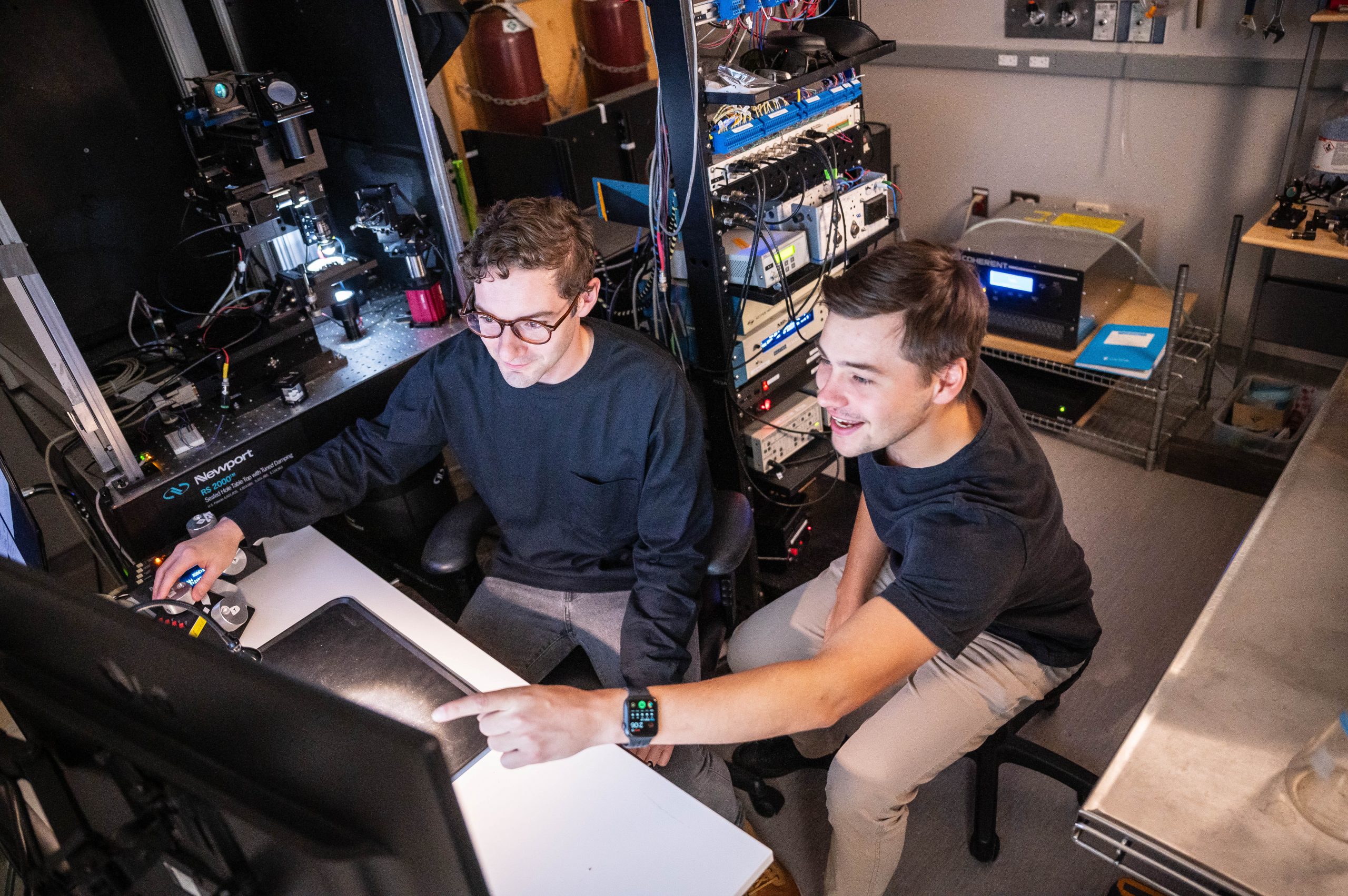
NINC supports several advanced light microscopy and imaging systems located in the Djavad Mowafaghian Centre for Brain Health including confocal and two photon microscopes as well as live cell imaging and optical coherence tomography.
For information on accessing these tools, please email Jeff LeDue (jledue@mail.ubc.ca)
Leica SP8 White Light Laser Confocal
Olympus FV1000 Confocal
Zeiss Live Cell Imaging System
Zeiss AxioZoom Macroscope
Zeiss 7MP FLIM Imaging System
Zeiss 7MP Dual Scan System
Thorlabs Telesto II: Optical Coherence Tomography Imaging System
Access to iMAP equipment is restricted to trained users from the iMAP Project team. Due to the complexity and expense of iMAP equipment, this project has its own fee structure and oversight committee. If your lab is interested in joining the iMAP Project please fill out this intake form
Fiber Photometry Instruments
Lattice Lightsheet Microscopy
Wide Field Multiphoton
Deep Multiphoton
Functional UltraSound Imaging (fUSI)